Scientists target major bottleneck in structural biology
X-ray free-electron lasers make membrane structure determination easier.
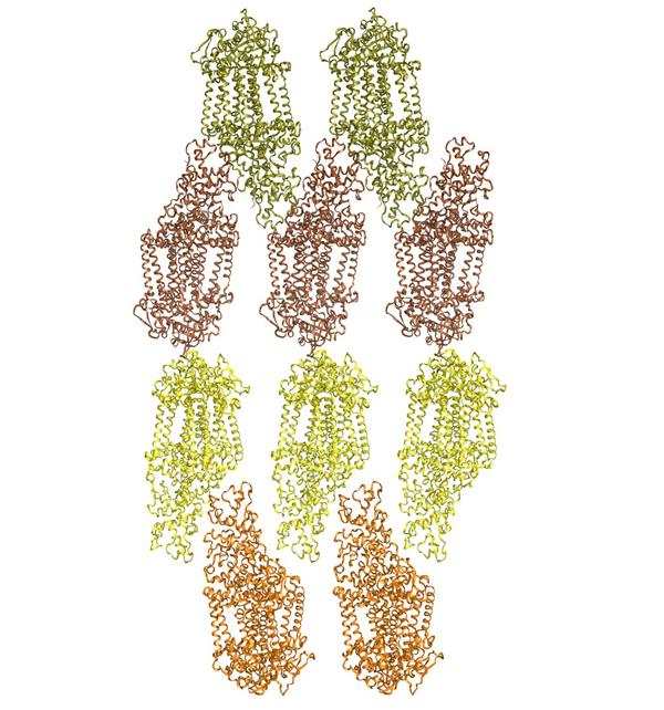
Crystal packing for the lipidic sponge phase microcrystals of Blastochloris viridis photosynthetic reaction center. Credit: Richard Neutze Group, Gothenburg University, Sweden
In terms of potential drug development, membrane proteins are of immense pharmaceutical interest. Studying their structures is an imperative step for successful drug design. Unfortunately, membrane protein structure determination remains a major obstacle in structural biology. An international research team, including DESY scientists, took a large leap forward in this field by analyzing microcrystals of a membrane protein embedded in lipids with an X-ray free-electron laser (X-FEL). This emerging method has the potential of rapidly increasing the number of available membrane protein structures, thereby assisting future drug design. The study has recently been published in “Nature Methods” (advance online 29.01.2012).
The promising results are attributable to the tremendous radiation power density of 600,000 trillion watts per square centimeter of the Linac Coherent Light Source (LCLS, SLAC National Accelerator Center, USA). The unparalleled intensity of X-FELs enabled the scientists to determine the structure of a photosynthetic reaction center from crystals that were approximately 100 times smaller than typically needed in conventional X-ray experiments. This approach exhibits remarkable progress in confronting the current challenge of obtaining large enough crystals in membrane structural biology.
Potential drug targets
All cells -the fundamental building blocks of living organisms -are surrounded by biological membranes, separating the cell interior from the environment. “Cell membranes regulate what comes into and goes out of the cell,” explains DESY scientist Henry Chapman, whose group at the Center for Free-Electron Laser Science (CFEL, Hamburg, Germany) participated in the study. Proteins associated with biological membranes are key players in the communication of cells with their environment. Receptor proteins on the outside of a cell, for instance, trigger changes inside the cell when binding signaling molecules such as hormones or neurotransmitters.
“In a lot of cases, membrane proteins are targets for drugs,” Chapman points out. “Caffeine, for example, works on a G protein-coupled receptor.” Membrane proteins not only affect coffee drinkers; they are of great interest for pharmaceutical research. Tailoring a new drug that effectively binds to a receptor protein is best achieved by first determining the structure of the receptor.
Notoriously difficult to crystallize
For many years, researchers have been successfully using X-ray crystallography to solve protein structures. When X-rays pass through a crystal, they scatter in specific directions and produce a so-called diffraction pattern. Scientists use this pattern to reconstruct the structure of the molecules inside the crystal. However, obtaining protein crystals of sufficient size is not always trivial. It is an endeavor that is particularly difficult for membrane proteins. The Stephen White group at the University of California in Irvine (USA) keeps track of the number of known membrane protein structures. As of mid-February 2012, out of the 73,492 protein structures deposited in the Protein Data Bank (PDB), a key resource in structural biology, only 320 are unique membrane proteins. Considering that approximately 30% of an organism’s proteins are thought to be associated with membranes, membrane proteins are dramatically under-represented in the PDB.
“Membrane proteins are so hard to crystallize because they need to sit in the membrane,” Chapman says. Biological membranes are made from lipids, a class of molecules that comprises fats and waxes. In most cases, removing the proteins from their “greasy” environment leaves them unstable. Therefore, one approach to crystallizing membrane proteins is to mimic their natural lipid environment.
Redefining crystal size requirements
In their study, researchers from Richard Neutze’s lab in Gothenburg (Sweden) crystallized a bacterial membrane protein from a lipidic sponge phase, named after the sponge-like shape of the lipid aggregates which form a stable mixture with the protein. Although the sponge phase allowed crystals to form, they were only a few thousandths of a millimeter in size. “At a synchrotron, we would need crystals about one hundred times larger because the strength of X-ray diffraction scales with the volume of the crystal,” Chapman explains. However, crystal size becomes less of an issue with X-FELs, the most intense X-ray sources to date. Reconstructing the protein structure from the diffraction images of many small crystals removes the bottleneck of obtaining a large enough crystal for synchrotron experiments.
At LCLS, the scientists injected a jet of lipidic sponge phase containing membrane protein crystals across a focused X-FEL beam. The X-ray pulses hitting the crystals were only 0.000 000 000 000 07 seconds (70 femtoseconds) short, but each of them produced an incredible 600,000 trillion watts per square centimeter on the sample. “Such a beam is powerful enough to burn through stainless steel,” Chapman says.
Using 265 individual diffraction patterns from randomly oriented microcrystals, the researchers were able to reconstruct the structure of the photosynthetic reaction center and show the potential of their method. Although the resolution of the structure was rather low, the scientists were able to trace characteristic features of the structure such as transmembrane α helices.
What’s next? Chapman and his group plan on applying their method to many more challenging membrane proteins. Moreover, recent developments at LCLS will provide researchers with shorter X-ray wavelengths than previously available and promise future membrane protein structures in atomic detail. “Shorter wavelengths result in a better resolution because smaller waves can interact with smaller objects,” Chapman says. “Atomic resolution is crucial if you really want to understand how drugs work.”
Manuel Gnida