First X-ray laser simulation suite explores single-particle imaging
Virtual X-ray laser developed to more efficiently define how structural biology’s “Holy Grail” method could work
Imagine taking an atomic-resolution X-ray picture of any individual biomolecule, without growing crystals, which can be extremely time-consuming or, in some cases, even impossible. Called single-particle imaging, this idea is often referred to as the “Holy Grail” of structural biology that could open new possibilities for many fields, including drug discovery and environmental science. In an effort to better understand the exact parameters for imaging single particles, an international research collaboration led by a team of scientists at CFEL and European XFEL – and partly from CFEL – has created the first detailed computer simulation suite for an entire experiment at an X-ray free-electron laser facility from start to end. A description of how this software works and what its potential scope is has been published in the current issue of Scientific Reports.
The new software suite —known as simS2E—is a large step forward in modelling single-particle imaging experiments. Previously, simulations of X-ray research focused on just one step of an experiment in isolation. These earlier models tended to consider only ideal scenarios and not the actual, complex conditions of a real X-ray free-electron laser experiment. By contrast, simS2E integrates all steps of an experiment and makes each step affect all following ones. The more accurate model of the experiment helps pinpoint parameters that could lead to a successful image of a single particle. As there are only a handful of instruments in the world potentially capable of high-resolution single particle imaging, including the upcoming SPB/SFX instrument at the European XFEL, this simulation suite can help a scientist determine optimal settings ahead of time and make significantly more efficient use of the limited time available at these experiment stations.
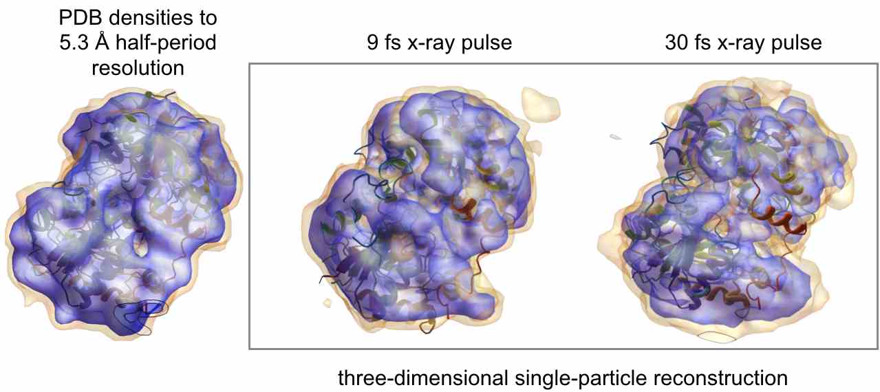
A simulated reconstruction of a molecule of a nitrogenase iron protein imaged using 9-fs long X-ray pulses (centre) versus 30-fs long pulses (right). (Credit: N. Duane Loh, National University of Singapore)
“What we’ve done is put together a comprehensive simulation pipeline, which simulates each step of a hypothetical single-particle imaging experiment, from source right through to analysis”, says European XFEL leading scientist Adrian Mancuso, who led the simS2E team. “We can ultimately give users more return on their time invested, since they could use this simulation to set up the best parameters possible for single-particle imaging—and hopefully the best data possible in the real experiment—in the least amount of time.” Already, the software has revealed useful information, confirming in its simulations theoretical expectations that shorter X-ray pulses will produce more easily interpreted data in these types of experiments.
To create simS2E—which stands for “simulation from source to experiment”—the team took some of the best existing simulations—designed at research centres around the world—of individual experiment steps such as X-ray generation from electrons and sample delivery, and made the output of each one into the input for the next. For the final part of the program, the team performed data analysis in the same way as planned for the SPB/SFX instrument. However, the team also designed the software suite in such a way that the individual simulations can be easily swapped with others, allowing the modelling to be tailored to other experiments, instruments, or even facilities, making simS2E potentially useful for other types of experiments aside from single-particle imaging.
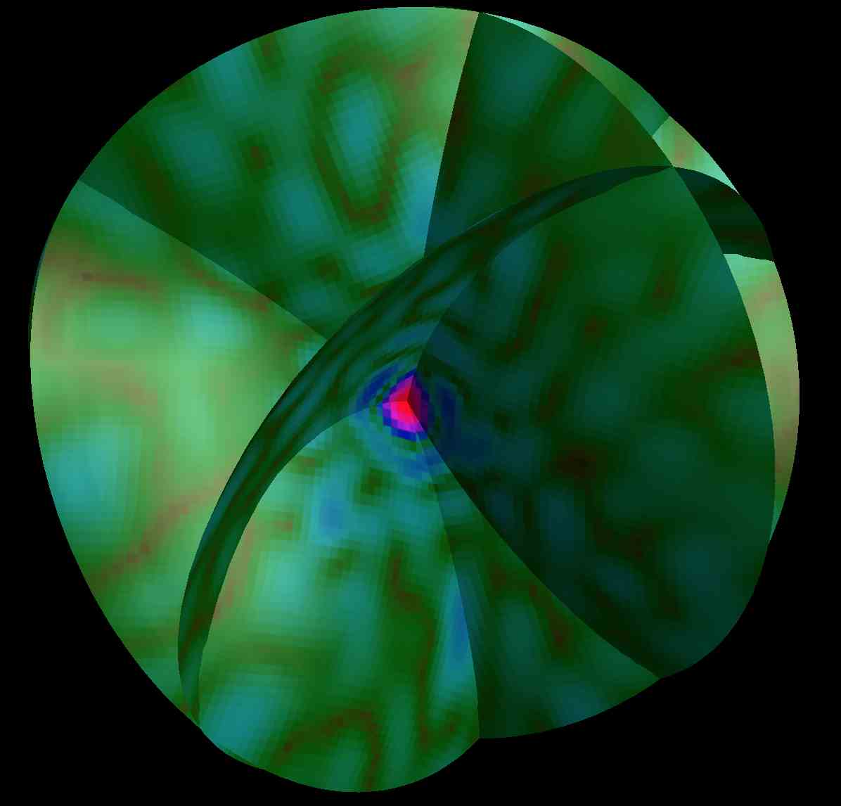
A representation of simS2E simulated two-dimensional diffraction patterns composed together into a three-dimensional pattern by the analysis software. (Credit: N. Duane Loh, National University of Singapore)
The research team included scientists from CFEL, the Hamburg Centre for Ultrafast Imaging (CUI), DESY, and European XFEL in Germany; SLAC National Accelerator Laboratory in the U.S.; Shubnikov Institute of Crystallography in Russia; IFJ-PAN in Poland; and the National University of Singapore. The simulation suite, which is part of a broader X-ray experiment simulation initiative led by Mancuso through the EU project EUCALL, is free to download at www.xfel.eu/simS2E.